
ItInsightsWorld AI Applications: How a Plain CT AI Flag Found an About 1.5 cm Lesion After Three Days of Dizziness
When an AI screening workflow examined a plain CT at the Ningbo University Affiliated People’s Hospital, it marked an approximately 1.5 cm lesion in the pancreatic region and raised the possibility of malignancy. The patient, Mr. Qiu, had come in with only three days of dizziness, not with a symptom pointing to the pancreas.
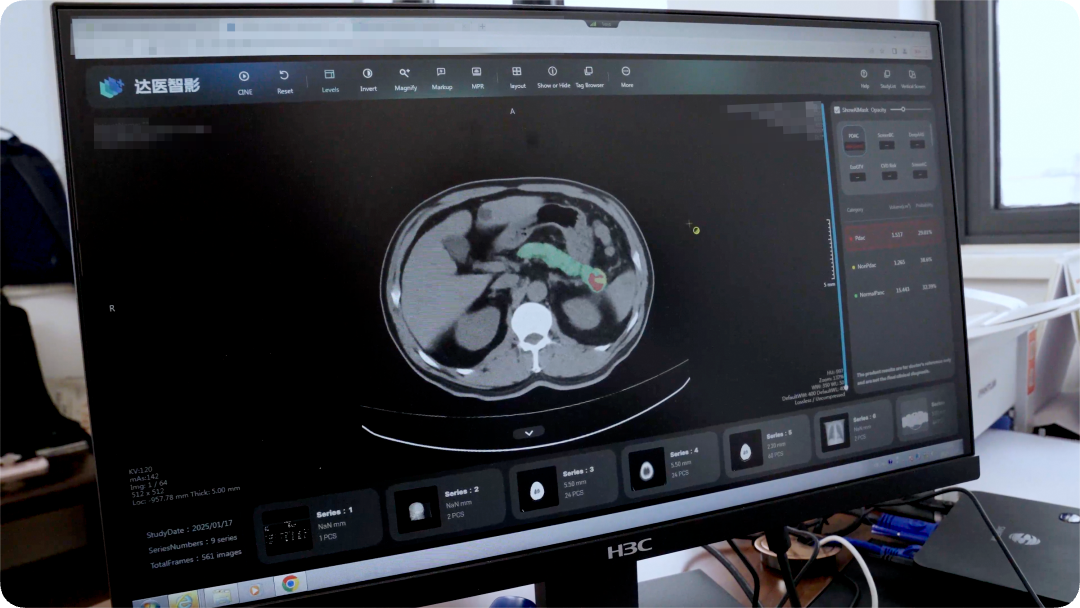
Case Anchor: A Chief Complaint That Did Not Match the Hidden Disease
Zhu Kelei, vice president and chief physician of hepatobiliary and pancreatic surgery, recalled the surprise: “This was something we doctors would not have imagined, because the patient’s chief complaint was dizziness, while pancreatic problems usually cause abdominal pain.”
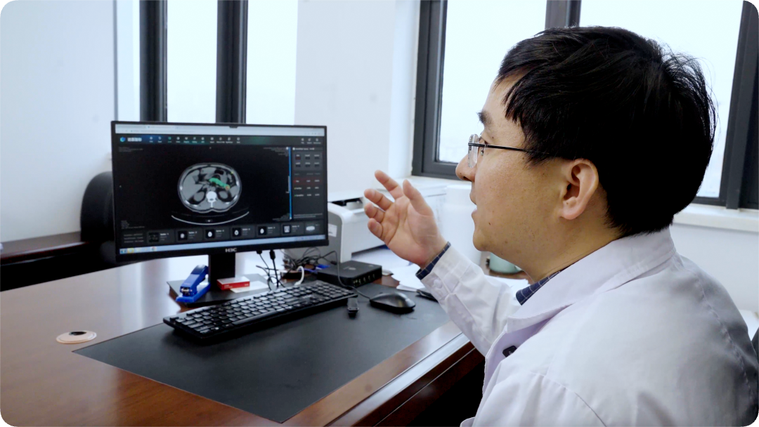
After the AI alert, clinicians moved quickly to enhanced CT, and the result pointed to pancreatic cancer. Mr. Qiu underwent surgery, and pathology confirmed pancreatic ductal adenocarcinoma. His daughter later said, “I feel so fortunate; my father really got his life back.”
Pancreatic cancer is often called the “king of cancers.” The organ sits deep in the abdomen, surrounded by the stomach, duodenum, and spleen, and early disease rarely produces clear symptoms. By the time discomfort is obvious, metastasis may already have happened.
Every year in China, more than 130,000 people are newly diagnosed with pancreatic cancer. More than 80% are already in late stage, and five-year survival is less than 10%.
How AI Applications Created a New Window
Since November 2024, the hospital has used medical AI from Alibaba DAMO Academy (Hupan Laboratory) and worked with Professor Jin Gang’s team at the Shanghai Pancreatic Disease Research Institute in a prospective clinical trial, becoming the first institution in Zhejiang Province to open a pancreatic cancer early screening clinic.
To date, the Ningbo project has screened CT imaging data from more than 40,000 people and found 8 pancreatic cancer cases, including 6 early-stage cases; all received surgical treatment.
The PANDA model study aligned enhanced CT images from more than 2,000 pancreatic cancer patients to plain CT images, so the system could learn subtle lesion patterns in lower-resolution scans.
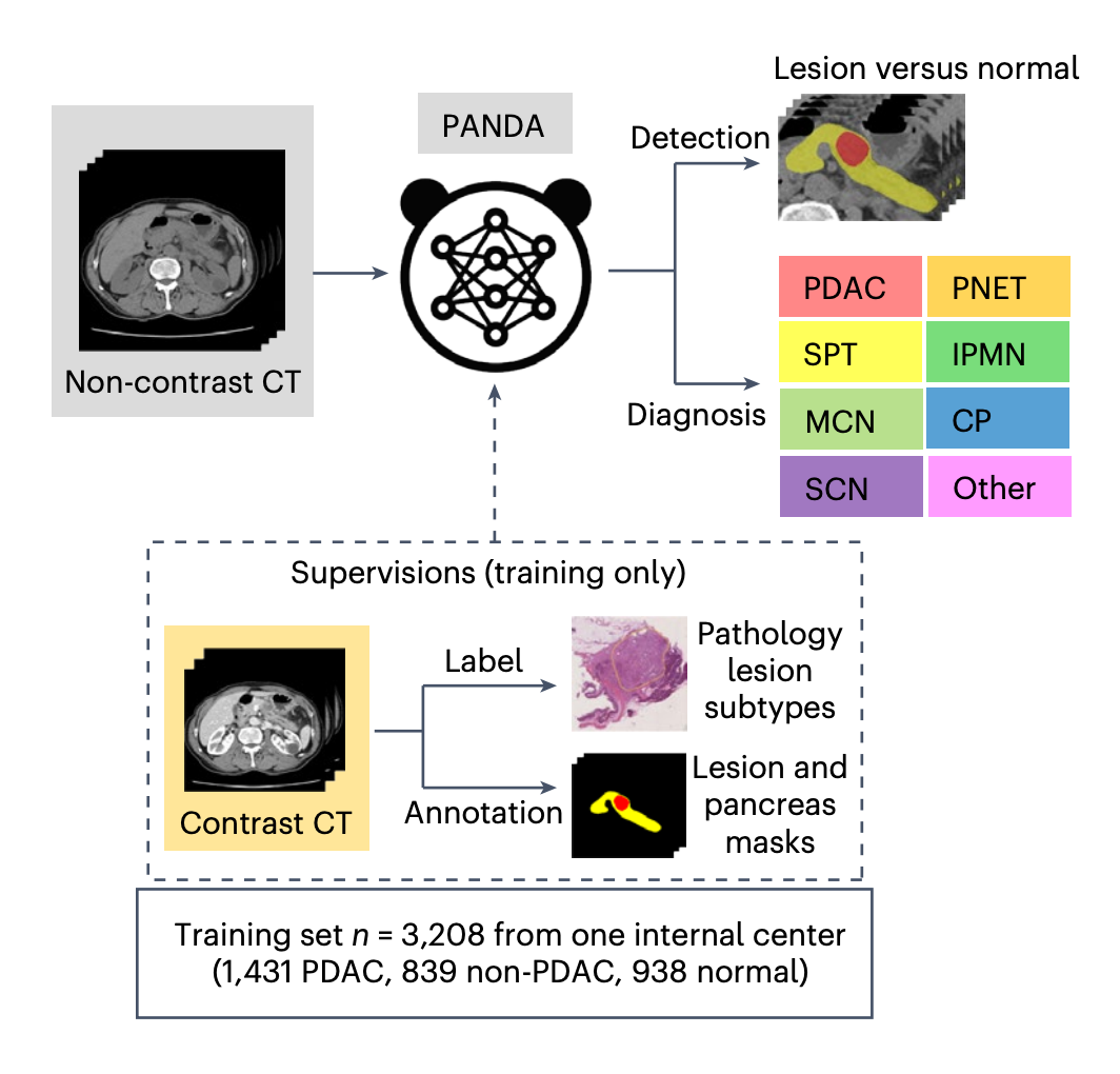
The reported performance was 92.9% sensitivity and 99.9% specificity, about 34% higher sensitivity than experienced imaging physicians.
Enhanced CT needs contrast injection and is costly, so it is not ideal for population-scale screening. Plain CT, by contrast, is already among the most common outpatient imaging tests. Opportunistic screening on these existing images means no extra exam burden for the patient: any routine visit or physical check-up can become an early cancer detection opportunity.
DAMO Academy says its multi-cancer early screening AI has begun global rollout, covering nine countries and regions and serving more than 20 million person-visits. The team is also pursuing a “one scan, multiple screenings” approach, where a single plain CT may help screen for pancreatic cancer, liver cancer, esophageal cancer, gastric cancer, colorectal cancer, and acute aortic syndromes.
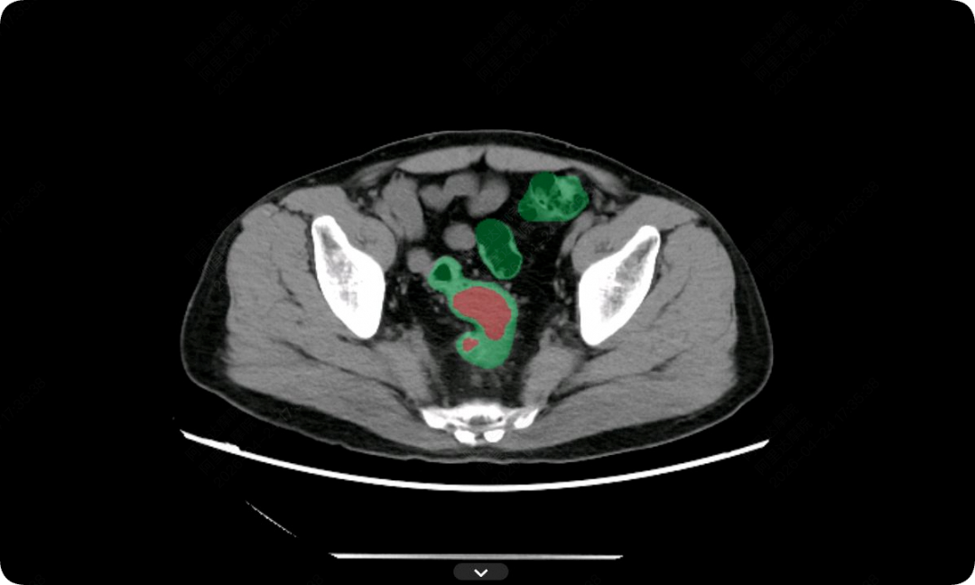
After the pancreatic cancer work, DAMO Academy released the intestinal cancer screening AI model DAMO COCA. Using plain CT images from 27,000 people, it accurately identified 5 previously missed colorectal cancer cases, with 86.6% sensitivity and 99.8% specificity.
Why This Matters for Application Builders
- Doctors remain irreplaceable in experience-based judgment and clinical decision making.
- Human attention can be pulled by the chief complaint, and fatigue affects reading of hundreds of dense image slices.
- AI does not get tired in the same way and is not misled by the patient’s presenting symptom, so it can surface lesions that might otherwise wait until late-stage symptoms appear.
Risk Prediction From Ordinary Photos
Recently, researchers at Harvard-affiliated Massachusetts General Hospital published work in Lancet Digital Health showing that a normal face photo could be used to predict a cancer patient’s death risk. The predictions were not fully accurate, but patients with higher predicted risk did show higher death probability.
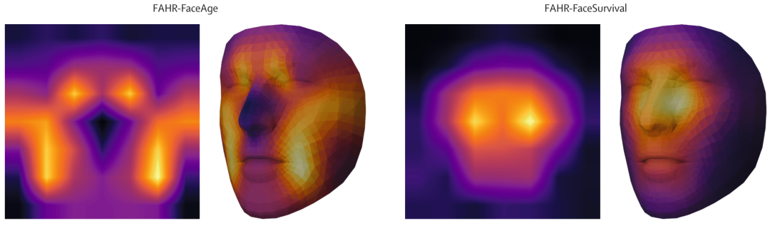
FaceAge uses the concept of biological age. Two people may both be 60 years old, yet one can look like 52 and the other like 71 because lifestyle, disease history, and genetics differ. Researcher Mak noted that smokers often look older than their actual age, and heavy drinkers may also appear older.
The team trained an algorithm to analyze 40 million ordinary face images without a task-specific label, then adapted the model to perform two jobs: estimating a person’s physiological age and predicting cancer patients’ survival odds.
For physiological age, AI relied on facial areas such as the nasolabial fold, sometimes described as the upper chin region, and parts of the forehead. For death-risk prediction, it focused more on the nose bridge and the area under the eyes.
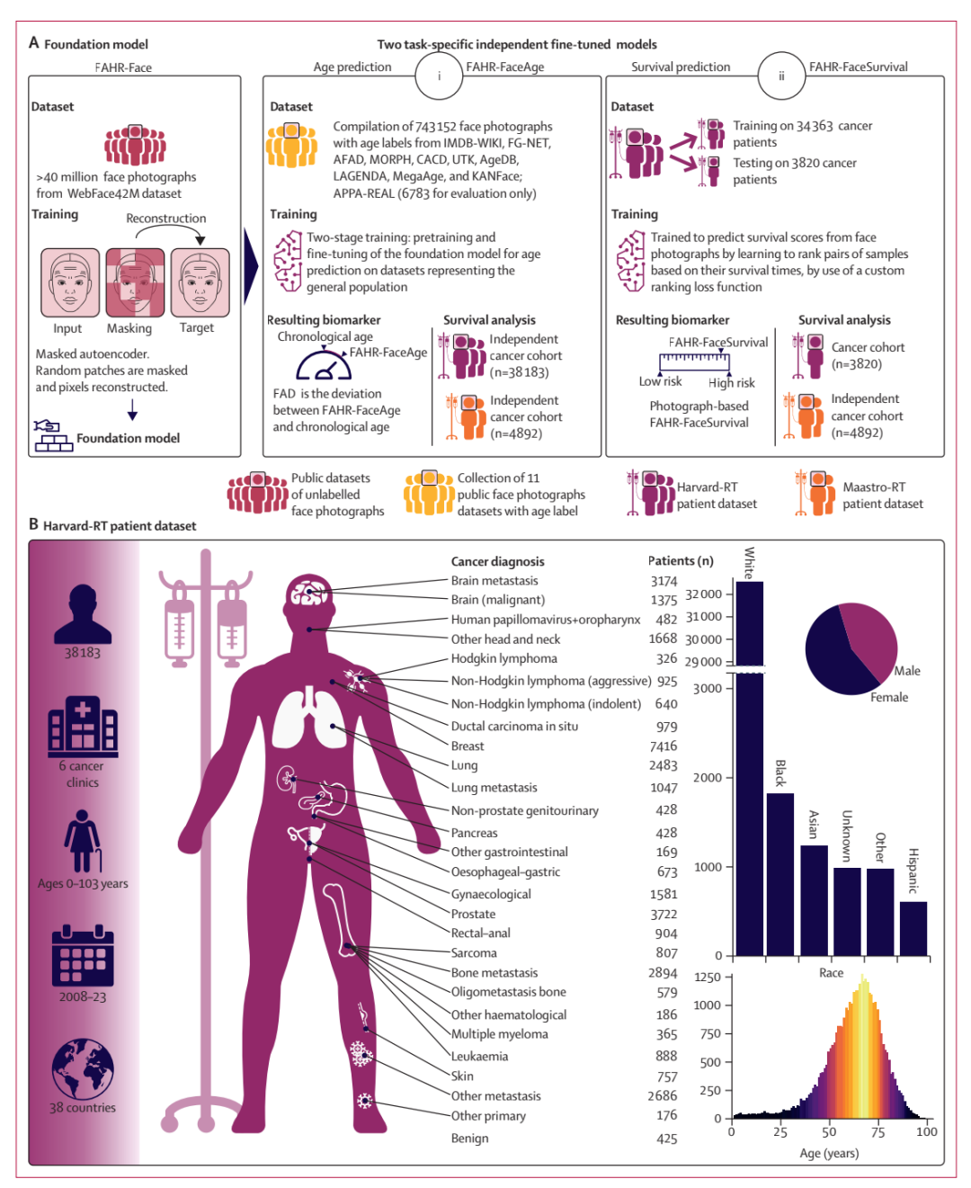
Whether the person was smiling or frowning, whether they were wearing makeup, and the lighting conditions did not materially affect age prediction. The AI could also account for common cosmetic surgery. One nuance was that people after rhinoplasty often looked slightly older in later photos than before the surgery.
For the death-risk task, in a test group of 3,820 cancer patients, more than half of those in the highest-risk score group died within 90 days after the photo was taken.
The researchers also examined lung cancer genetic data and found that FaceAge scores were negatively correlated with CDK6, a gene involved in cell-cycle regulation and linked to progression in several cancers. A model trained only on facial images had captured, at some level, a signal connected to genetic biology.
Sophont’s chief executive shared another application example involving osteosarcoma. The company said its Holon-S1 model analyzed pathology slides without specialized osteosarcoma training, extracting key biological information: fibroblast-related genes showed overexpression.
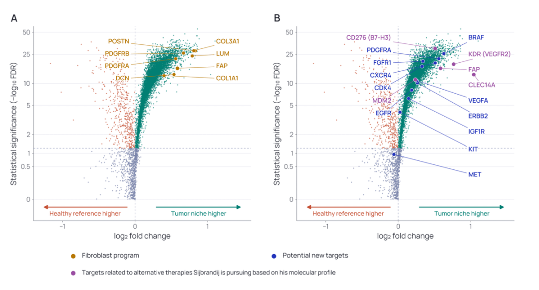
The patient’s medical team had already found the same overexpression through advanced molecular testing, and had identified a corresponding treatment target.
Governance: The Harder Problem Behind the Model
FaceAge’s authors raised a worry: the tool could assist clinicians, but it might also attract life insurers or employers who want to estimate personal risk. The moment Mr. Qiu was pulled from an ordinary queue by an incidental AI flag was a case of technology used for a beneficial purpose. A future insurance applicant flagged as high risk from a photo would create a different question.
The visual features of aging learned by a model may not apply evenly across all skin colors and facial structures. If the system systematically overestimates biological age for certain ethnic groups, that error could quietly reproduce and amplify existing inequality, and the people affected may not know why they were misjudged.
Earlier research found that some commercial models used to predict healthcare needs systematically underestimated the needs of Black patients because historical medical spending served as a proxy for health, while Black patients with the same condition had historically received fewer medical resources.
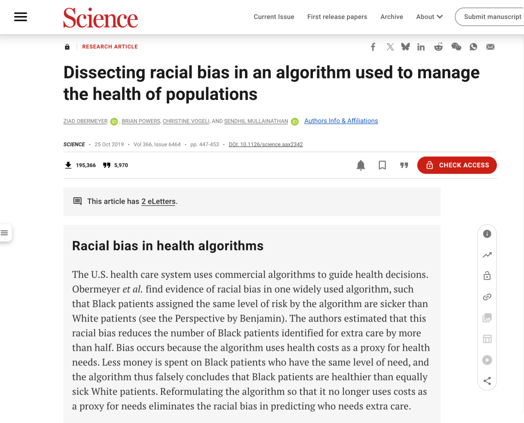
Researchers emphasize that FaceAge must be tested in more diverse populations before clinical use, with potential errors identified and removed. The harder task is not training the model, but handling the social, legal, and fairness questions that follow.
Comparison for AI Applications Teams
| Screening path | AI on existing plain CT | Enhanced CT after suspicion |
|---|---|---|
| Patient burden | No extra exam if the scan is already made | Additional contrast injection and visit cost |
| Use case | Opportunistic detection when symptoms do not match the disease | Confirm or characterize a suspected lesion |
| Workflow fit | Can review large volumes of routine images | Better once a clinical question is known |
| Failure point | Missed if routine images are not routed to screening | Missed if clinicians do not suspect pancreatic disease |
Step-by-Step: From Application Signal to Clinical Action
- Connect AI to images already produced in routine care, such as plain CT.
- Let the model flag regions that do not match the presenting symptom, such as an approximately 1.5 cm pancreatic lesion after only three days of dizziness.
- Escalate to a higher-resolution diagnostic step, such as enhanced CT.
- Confirm with clinical review and pathology, as in pancreatic ductal adenocarcinoma.
- Feed outcomes back into screening metrics, including number of scans, cases found, early-stage share, sensitivity, specificity, and false-positive review.
Checklist for Evaluating a Health AI Workflow
- Is the target disease hidden and low-symptom, such as pancreatic cancer?
- Can the screening use existing images instead of creating new patient burden?
- Does the AI reduce human blind spots caused by fatigue and symptom anchoring?
- Are follow-up pathways, clinician review, and pathology confirmation defined?
- Is performance tested across diverse populations, not only overall averages?
FAQ
Can AI find cancer when the chief complaint is unrelated?
Yes. In the Ningbo University Affiliated People’s Hospital case, AI found an approximately 1.5 cm pancreatic lesion in a patient whose only reported symptom was three days of dizziness. Enhanced CT and pathology then confirmed pancreatic ductal adenocarcinoma.
Does high sensitivity mean the model replaces doctors?
No. The model reached 92.9% sensitivity and 99.9% specificity, but the case remained a clinical pathway: AI alerted, physicians ordered enhanced CT, and pathology confirmed the diagnosis. Doctors remain irreplaceable for judgment and treatment decisions.
What is opportunistic screening?
It is screening that happens on data already collected for another purpose. With plain CT, a routine scan can trigger an early cancer alert without requiring an extra exam, which is especially important when diseases are silent early.
Why do facial-photo risk models need caution?
FaceAge may capture biological aging and risk signals, but it can be misused by insurers or employers. If visual aging cues vary by skin color or facial structure, the model may systematically overestimate risk for some groups and hide that error from patients.
Common Pitfalls in Applying This Kind of AI
- Treating an AI alert as a diagnosis rather than a trigger for confirmatory workup.
- Assuming symptoms must match the disease before screening.
- Reporting sensitivity and specificity without subgroup validation.
- Deploying risk-prediction models before defining who may see or act on their outputs.
- Using proxy labels, such as historical spending, without auditing structural bias.
Action Recommendations for Product and Operations Leaders
- Start where images already exist, not where new data collection is required.
- Choose application areas where the problem is silent but consequential, such as early cancer detection.
- Design escalation rules before deploying alerts, including who reviews, who orders enhanced CT, and how outcomes are tracked.
- Measure both clinical impact and equity impact, including early-stage rate, missed cases, subgroup performance, and user burden.
- Keep governance close to the model: fairness review, consent boundaries, and limits on insurer or employer use should be part of the launch checklist.
Bottom Line
For ItInsightsWorld AI Applications readers, the value is not simply that an algorithm outperformed a radiologist. It is that application-layer AI can change outcomes when it is embedded in existing workflows, catches mismatches between patient stories and disease reality, and routes people into timely clinical confirmation. The same embedding logic also explains the concern about facial-photo risk models: once a system is in the workflow, its outputs can shape access, cost, and diagnosis. The harder challenge after model training is ownership, fairness, and responsible deployment.